|
 |
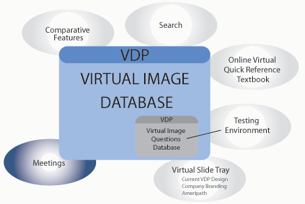
| VDP
|
To emulate the examination of glass slides on a conventional light microscope by creating an online "virtual microscope".
|
|
|
|
|
A virtual microscope application designed to enhance the educational experience of Dermatology residents. It uses streaming imaging techniques that enable educators to duplicate the experience of using a light microscope. This tool is critical to the learning process in the field of dermatopathology, for with it, clinical and histopathological information for almost any disease process is only a mouse click away.
The Internet has become one of the most widely used tools in medical education. Much of its popularity stems from its ability to blend images with text in an interactive manner. Today, clinical and histopathological information for almost any disease process is only a mouse click away, and newer techniques for dynamic image display are revolutionizing the field of dermatopathology education. These streaming imaging techniques enable educators to incorporate images that duplicate the experience of using a light microscope. This virtual microscope emulation is critical to the learning process, compelling the user to not only interpret images, but to select the fields of interest necessary to correctly diagnose a tissue specimen. Derm.md has developed a novel way to teach dermatopathology using "zoomable" imaging via the Internet to enhance the educational experience.
Using an ultra-fast proprietary microscope slide scanner that digitizes an entire slide at high resolution in minutes, imagery data is captured at approximately 3 GB per minute. Following wavelet compression, a typical slide is reduced in size to 150 MB. The typical scanning resolution is 54,000 pixels per inch, with higher resolutions possible. Images are delivered over the web in a proprietary format, which is conceptually a pyramidal stack of jpeg-compressed images with a full sized, full resolution image at the base and a small, low-resolution image at the top. A zoom function jumps from one layer in the stack to the next, changing magnification with the click of the mouse. This supercedes previous attainable resolutions and more accurately emulates a conventional light microscope.
"glass slide specimens only a click away"
Our goal in the creation of this unique concept for teaching Dermatopathology was to emulate the examination of glass slides on a conventional microscope by creating a complete online learning environment.
The virtual slide specimen is a digitally acquired image delivered over the web in a novel format. Conceptually, this slide is made up of a pyramidal stack of compressed images, with an extremely large, high-resolution image at the bottom of the pyramid. Starting at the tip of the pyramid, or the lowest power, the user can obtain higher and higher magnifications to recognize histologic patterns of cutaneous diseases and evaluate precise cellular detail and morphology. In short, the more the image is magnified, the more detailed information the user gains about the image. One may select from any number of diagnoses and view each at one’s leisure.
Most importantly, the user is in control, achieving a degree of interactivity that compels them to select fields of view necessary to make a diagnosis, as opposed to being guided towards a diagnosis by examination of a series of pre-selected pertinent images.
Virtual Dermpath is provided at no charge to Dermatology residents and fellows in training. If you would like to get started with VDP, register at: virtualdp.com
|
|
|
|